Lipid nanoparticles are currently the most widely used non-viral system for delivering RNA into cells, enabling cytosolic release and transient protein expression. Their apparent effectiveness depends less on the mRNA sequence itself than on lipid composition, particle attributes, and the biological context in which delivery occurs. For academic researchers, understanding these constraints is essential to designing experiments that are interpretable, reproducible, and biologically meaningful.
This page is written for researchers using or evaluating lipid nanoparticles for RNA delivery in experimental systems. It focuses on biological mechanisms, experimental constraints, and common failure modes.
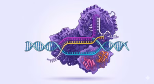
What lipid nanoparticles do in RNA delivery
Lipid nanoparticles are nanoscale lipid assemblies designed to encapsulate nucleic acids and facilitate their entry into cells.
In RNA applications, their primary role is to protect RNA from extracellular degradation, promote cellular uptake via endocytosis, and enable partial release of RNA into the cytosol, where translation occurs.
In practice, encapsulation efficiency alone does not predict performance. Delivery outcomes are shaped by how particles interact with biological fluids, how cells internalize them, and how efficiently RNA escapes endosomal compartments.
These factors vary across cell types and experimental systems, which explains why identical formulations can behave differently in different contexts. For example, if mRNA expression varies unexpectedly across models, delivery rather than sequence design is often the first variable to examine.

LNP composition influences experimental outcomes
Most LNP formulations used for RNA delivery rely on four functional lipid components: ionizable lipids, helper lipids, cholesterol, and PEG-lipids. Each contribute differently to particle behavior, cellular uptake, and intracellular RNA.
Ionizable lipids
They bind RNA during formulation and trigger its release inside cells. Because they carry a positive charge at low pH, they interact electrostatically with negatively charged RNA during particle assembly. Once inside the cell, as the endosome acidifies to around pH 5.0–5.5, the same charge response destabilizes the endosomal membrane and allows RNA to partially escape into the cytosol. The pKa of the ionizable lipid, optimally between 6.2 and 6.8, determines how well it balances these two demands: neutral enough in the bloodstream to avoid toxicity, reactive enough inside the endosome to enable release. The structure of the lipid tail separately governs how quickly the lipid degrades, which affects both how long the effect lasts and how well the formulation is tolerated in vivo.
Helper lipids
Helper lipids define how the particle membrane is organized and how it behaves inside cells. The two most common choices, DSPC and DOPE, produce meaningfully different outcomes. DSPC forms a stable, ordered membrane layer that protects the particle during circulation. DOPE, under the acidic conditions of the endosome, shifts into a cone-shaped geometry that disrupts the endosomal membrane and improves RNA release into the cytosol. Choosing between them is therefore a deliberate formulation decision, not a substitution.
Cholesterol
Cholesterol keeps the particle membrane fluid and structurally stable, which matters both during storage and after the particle enters the cell. It also influences how the lipid mixture behaves during endosomal escape, as its membrane-condensing effect modulates the overall fusogenic response of the formulation.
PEG-lipids
They coat the particle surface with a hydrophilic layer that controls size, prevents aggregation, and slows clearance by shielding the particle from immune recognition. The trade-off is that the same coating can block cellular uptake and make endosomal escape harder by stabilizing the membrane against disruption. For this reason, PEG-lipid content, typically kept between 1–2.5% in clinical formulations, needs to be matched to the delivery route and target cell type rather than set as a fixed parameter
These four components interact rather than act independently, small changes in lipid ratio, grade, or identity can produce disproportionately large effects on biological outcome. Any compositional modification should be treated as a new experimental condition, not a minor optimization.
Why mRNA delivery remains biologically constrained
What happens to LNP–mRNA complexes inside cells
Following exposure to cells, lipid nanoparticles are taken up primarily through endocytosis. As endosomes acidify, ionizable lipids within the particle become protonated, destabilizing the endosomal membrane and enabling limited release of mRNA into the cytosol.
This release step is the principal bottleneck in mRNA delivery. Only a small fraction of internalized mRNA typically reaches the translational machinery, and increasing dose does not produce proportional increases in protein expression. For this reason, apparent transfection efficiency often reflects a convolution of delivery, translation, and protein stability rather than direct mRNA availability.
That is why when optimizing dose, it should be considered whether increases in signal reflect improved delivery or simply downstream amplification.
Biodistribution and intracellular limitations
Unmodified lipid nanoparticles often accumulate preferentially in the liver due to interactions with serum proteins and hepatic uptake mechanisms. While this behavior can be advantageous in some contexts, it limits applicability to other tissues.
Even after uptake, endosomal trapping remains a dominant limitation. Adjusting lipid composition or incorporating fusogenic elements can improve release efficiency, but no current approach eliminates this bottleneck entirely.
Consequently, delivery efficiency often defines both dose requirements and observed biological effect
What determines whether an LNP works in a given model
Successful mRNA delivery depends on interacting variables that are often underestimated in academic research.
- Cell type is a major determinant, as uptake pathways, endosomal maturation, and innate immune sensing differ widely between immortalized cell lines, primary cells, and tissues.
- mRNA design also matters, including length, untranslated regions, secondary structure, and nucleotide modifications, all of which influence stability, translation, and immune activation. Particle attributes such as size, surface chemistry, and charge affect uptake and trafficking independently of encapsulation efficiency.
- Assay design influences interpretation, as reporter systems can mask delivery limitations through nonlinear signal amplification.
Together, these factors explain why formulations that appear robust in one system may fail in another.
Common experimental failure modes in mRNA–LNP studies
Several recurring patterns reduce interpretability in LNP-based mRNA experiments.
- Reporter expression is frequently overinterpreted without accounting for protein half-life or amplification effects. Delivery success is often inferred from transfection efficiency, even though most internalized mRNA never reaches ribosomes.
- Batch-to-batch variability in LNP preparation can introduce uncontrolled noise, particularly when formulation parameters are adjusted informally.
- mRNA constructs are sometimes changed without re-optimizing formulation, despite strong sequence dependence on delivery efficiency.
Recognizing these failure modes early improves reproducibility and reduces the risk of false mechanistic conclusions.
When off-the-shelf systems stop being sufficient
Commercial transfection reagents and generic LNP formulations are valuable for early proof-of-concept work. However, as research questions become more biology-specific, these systems often fail to provide consistent or interpretable results.
Custom formulation becomes relevant when delivery efficiency varies strongly across models, when immune activation interferes with readouts, or when reproducibility becomes a limiting factor. At this stage, delivery should be treated as an experimental parameter rather than a fixed input.
Comparison of common mRNA delivery approaches
The table below compares commonly used mRNA delivery approaches by intracellular destination, duration of expression, and experimental flexibility to support method selection.
| Delivery approach | Strengths | Limitations | Expression window |
|---|---|---|---|
|
Lipid nanoparticles (ionizable LNPs) |
Scalable in vivo delivery; clinically validated for mRNA |
Endosomal escape is inefficient (<2–5% estimated); liver tropism after systemic dosing |
24–96 h (context-dependent) |
|
Targeted LNPs (tLNPs) |
Improved cell or tissue selectivity; enables non-hepatic targeting in vivo; adaptable chemistry |
Low endosomal space. Performance depends on receptor density and internalization rate. Complex formulation |
24–96 h |
|
Cationic lipofection reagents |
High efficiency in immortalized cell lines |
Cytotoxicity; poor performance in primary cells; not suitable for systemic in vivo delivery |
24–72 h |
|
Polymeric nanoparticles (e.g., PEI-based) |
Customizable chemistry |
High toxicity; batch variability; limited clinical translation for mRNA |
24–72 h |
Conclusion on LNP-based RNA delivery
Lipid nanoparticles enable mRNA, siRNA, circRNA and saRNA delivery by protecting RNA, promoting cellular uptake, and enabling limited cytosolic release. Their effectiveness is governed primarily by formulation variables and biological context rather than by mRNA sequence alone. Treating delivery as a central experimental constraint improves reproducibility, interpretability, and downstream relevance of mRNA research.
Frequently asked questions
What are lipid nanoparticles used for in mRNA research?
Lipid nanoparticles are used to deliver mRNA into cells by protecting it from degradation, enabling cellular uptake, and allowing partial release into the cytosol where translation occurs. In research settings, they are commonly used to study protein expression, gene function, and RNA-based perturbations both in vitro and in vivo.
Why are lipid nanoparticles preferred over viral vectors for mRNA delivery?
Lipid nanoparticles deliver mRNA directly to the cytosol without requiring nuclear entry or transcription. This enables transient, dose-controlled expression and avoids risks associated with genomic integration and long-term persistence that are inherent to DNA-based viral vectors.
Where does LNP-delivered mRNA act inside the cell?
LNP-delivered mRNA acts in the cytosol, where it is translated by ribosomes into protein. The nucleus is not involved in the mechanism of action, and nuclear entry is neither required nor desired for mRNA therapeutics.
What is the main limitation of lipid nanoparticles for RNA delivery?
The primary limitation is inefficient endosomal escape. Although LNPs are readily taken up by cells, only a small fraction of internalized RNA typically reaches the cytosol. This bottleneck often defines dose requirements and limits the efficiency of protein expression.
Why does mRNA expression not scale linearly with LNP dose?
Increases in dose often lead to higher cellular uptake but not proportionally higher cytosolic delivery. Endosomal trapping, saturation of escape mechanisms, and innate immune activation can all limit the fraction of mRNA that is translated, resulting in nonlinear dose–response behavior.
Do all cell types respond similarly to LNP-mediated RNA delivery?
No. Uptake pathways, endosomal processing, and innate immune sensing differ substantially across cell types, primary cells, and tissues. As a result, an LNP formulation that performs well in one model may fail or behave unpredictably in another.
Can RNA sequence changes affect LNP delivery performance?
Yes. mRNA length, secondary structure, untranslated regions, and nucleotide modifications can influence stability, immune recognition, and translation efficiency. These changes can alter apparent delivery performance even when the LNP formulation remains unchanged.
When are commercial transfection reagents no longer sufficient?
Commercial reagents are often adequate for early proof-of-concept experiments. They become limiting when delivery efficiency varies strongly across models, when immune activation interferes with readouts, or when reproducibility becomes a critical constraint. At that point, delivery should be treated as an experimental variable.
Are lipid nanoparticles mainly liver-targeted?
Unmodified LNPs often accumulate preferentially in the liver due to interactions with serum proteins and hepatic uptake mechanisms. While this can be advantageous for certain applications, it limits delivery to other tissues unless formulations are specifically engineered.
How should academic researchers think about translation when using LNPs?
Even in basic research, delivery assumptions can propagate downstream. Parameters that appear inconsequential in vitro, such as particle size or lipid ratios, often become critical in vivo. Designing experiments with delivery constraints in mind improves interpretability beyond the immediate study.
References
- Kulkarni JA, et al. The current landscape of nucleic acid therapeutics. Nature Nanotechnology. 2021. https://www.nature.com/articles/s41565-021-00898-0
- Jayaraman M, et al. Maximizing the potency of siRNA lipid nanoparticles for hepatic gene silencing in vivo. Angewandte Chemie. 2012. https://doi.org/10.1002/anie.201203263
- Semple SC, et al. Rational design of cationic lipids for siRNA delivery. Nature Biotechnology. 2010. https://www.nature.com/articles/nbt.1602
- Sabnis S, et al. A novel amino lipid series for mRNA delivery. Molecular Therapy. 2018. https://doi.org/10.1016/j.ymthe.2018.03.010
- Koynova R, Tenchov B. Lipid phases and lipid phase transitions of relevance to drug delivery. OA Biochemistry. 2013. https://www.ncbi.nlm.nih.gov/pmc/articles/PMC4314312/
- Patel S, et al. Naturally-occurring cholesterol analogues in lipid nanoparticles induce polymorphic shape and enhance intracellular delivery of mRNA. Nature Communications. 2020. https://www.nature.com/articles/s41467-020-14527-2
- Mui BL, et al. Influence of polyethylene glycol lipid desorption rates on pharmacokinetics and pharmacodynamics of siRNA lipid nanoparticles. Molecular Therapy Nucleic Acids. 2013. https://doi.org/10.1038/mtna.2013.66
- Akinc A, et al. The Onpattro story and the clinical translation of nanomedicines containing nucleic acid-based drugs. Nature Nanotechnology. 2019. https://www.nature.com/articles/s41565-019-0591-y
- Gillmore R, et al. CRISPR-Cas9 in vivo gene editing for transthyretin amyloidosis. New England Journal of Medicine. 2021. https://doi.org/10.1056/NEJMoa2107454
- Kauffman KJ, et al. Optimization of lipid nanoparticle formulations for mRNA delivery in vivo with fractional factorial and definitive screening designs. Nano Letters. 2015. https://doi.org/10.1021/acs.nanolett.5b02497
- Schoenmaker L, et al. mRNA-lipid nanoparticle COVID-19 vaccines: Structure and stability. International Journal of Pharmaceutics. 2021. https://doi.org/10.1016/j.ijpharm.2021.120586
- Wittrup A, Bhatt DL. Endosomal escape and the knockdown efficiency of siRNA nanoparticles. Nature Biotechnology. 2015. https://www.nature.com/articles/nbt.3330